










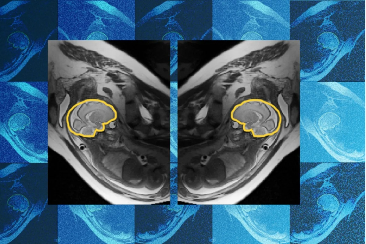
The development of the human brain is a marvel of biological precision, yet it remains one of the most vulnerable processes in early life. From the first weeks of gestation through the months following birth, the brain undergoes a radical transformation, building the complex architecture that will underpin a lifetime of cognition. However, this period of rapid growth is susceptible to genetic, environmental, and metabolic disruptions. When these disruptions occur, early detection is paramount. Yet, for decades, clinicians have been hampered by a stubborn physical reality: you cannot ask a fetus to sit still.
A groundbreaking collaboration between researchers at MIT, Boston Children’s Hospital, and the French National Institute for Research in Digital Science and Technology (Inria) has introduced a potential paradigm shift in prenatal diagnostics. The team has developed "E(3)-Pose," a deep learning model that utilizes physics-informed artificial intelligence to track fetal movement in real-time during an MRI scan. By enabling the capture of high-resolution images despite constant fetal motion, this technology promises to transform how radiologists diagnose developmental abnormalities, moving the field toward a future of faster, more accurate, and more comfortable prenatal care.
The Challenge of the Moving Target
In standard clinical practice, a routine prenatal ultrasound acts as the first line of defense. If a sonographer identifies a potential brain abnormality, the patient is typically referred for a fetal magnetic resonance imaging (MRI) scan to get a clearer, more detailed view. However, fetal MRI is notoriously difficult.
To produce a high-resolution 3D volumetric image, the MRI machine must capture a series of 2D "slices" of the fetal head. These slices are then reconstructed into a single, cohesive image. The problem arises when the fetus shifts position during the procedure. Because the slices are captured sequentially, a movement halfway through the scan can misalign the entire data set. This results in "gaps" in the coverage or distorted slice orientations, leaving radiologists with incomplete or blurry data.
"One of the problems with having the baby move is that the slices are all over the place," explains Ramya Muthukrishnan, a PhD student at MIT and the first author of the research. These gaps are not merely aesthetic issues; they are clinical hazards. "There are very fine details that you need a very good volumetric picture of the brain to see," Muthukrishnan notes. "If you have coverage gaps, you can’t see critical regions, and you need to be able to see well to detect abnormal development."
Chronology of Innovation: From Frustration to Physics
The inception of E(3)-Pose was born out of clinical empathy. Early in her doctoral studies, Muthukrishnan spent time shadowing radiologists at Boston Children’s Hospital. She observed a recurring, distressing scenario: pregnant patients enduring an hour-long, uncomfortable MRI procedure, only for the scan to be aborted due to excessive fetal movement. The result was often a failure to obtain the diagnostic-quality imagery necessary for doctors to make informed decisions.
"Seeing frustration about it helped me decide what I wanted to do with my PhD, because it felt like this was a pain point," she recalls.
The development process was anything but linear. As a computer scientist by training, Muthukrishnan found herself thrust into the world of hardware-level MRI physics. The transition was arduous. "I was surprised when I had to go and implement this on the scanner," she says. "I was not prepared for how many times I would crash the scanner because putting things on hardware and getting it to work on hardware is a different beast than training a model to run on data on a GPU."
The team’s breakthrough came when they decided to move beyond traditional "black box" machine learning. Instead of simply training a model to recognize patterns in raw data, they embedded the actual laws of physics into the neural network. By incorporating the inherent left-right symmetry of the human head into the model’s architecture, E(3)-Pose gained an innate understanding of the subject’s orientation. This "physics-informed" approach allows the model to predict and adjust for motion far more effectively than previous iterations.
Throughout this process, the team leaned on interdisciplinary support. Borjan Gagoski, an assistant professor of radiology at Harvard Medical School, developed specialized imaging sequences that allowed for the high-frequency data acquisition needed to train the model. Aryn Lee, a research assistant at Boston Children’s, managed the recruitment of pregnant volunteers, ensuring the model was trained on real-world clinical data.
Supporting Data and Technical Architecture
The technical core of the E(3)-Pose model lies in its use of "equivariance"—a geometric concept that allows the neural network to understand how the orientation of an object changes in space. By baking this into the architecture, the model does not have to "learn" that a brain is the same object when it turns; it fundamentally understands that the spatial relationship of the brain’s structures remains constant, even if the brain itself has rotated within the field of view of the MRI.
The research, presented at the 2026 European Conference on Computer Vision, highlighted several key performance metrics:
- Motion Robustness: The model demonstrated an unprecedented ability to reconstruct high-resolution volumes from noisy, motion-corrupted 2D slices.
- Physics Integration: Unlike standard convolutional neural networks, E(3)-Pose requires significantly less training data because it relies on the geometric constraints of the human anatomy.
- Clinical Efficacy: Preliminary testing suggests that the time required for a successful scan can be reduced, significantly lowering the burden on the patient and increasing the throughput of the radiology suite.
The project was guided by a heavy-hitting panel of experts, including Polina Golland, a principal investigator at the MIT Jameel Clinic and CSAIL; Elfar Adalsteinsson, an MIT professor of electrical engineering and computer science; and Ellen Grant, a leader in pediatric radiology at Harvard Medical School. Benjamin Billot of Inria provided the foundational methods that allowed the project to move from concept to implementation.
Official Responses and Clinical Impact
The implications of this work have drawn high praise from the medical and academic communities. "Ramya’s work elegantly combines geometric concepts like equivariance and symmetry with the power of deep learning to change how MRI scanners acquire the images," says Polina Golland. "It’s an impressive research project that is deep and fundamental in how it models image acquisition, with clear clinical impact."
Dr. Ellen Grant, who directs the Fetal Neonatal Neuroimaging and Developmental Science Center (FNNDSC) at Boston Children’s Hospital, believes the technology will be transformative for the field. "Muthukrishnan’s work will automate fetal brain MRI, replacing today’s slow, manual process with faster and more comfortable scans for mothers, while also providing higher-quality images for radiologists to interpret," Grant stated.
By reducing the frequency of re-scans and aborted procedures, E(3)-Pose could effectively lower the cost of diagnostic care while simultaneously increasing the volume of patients that clinics can serve, ensuring that fewer high-risk pregnancies go without the necessary diagnostic oversight.
Broader Implications: Beyond the Fetal Brain
While the immediate focus of E(3)-Pose is the fetal brain, the potential applications for this technology are vast. The model’s ability to track objects through noisy, 3D environments has caught the attention of researchers in other fields.
- General Medical Imaging: The principles applied to the fetal head could be adapted for other hard-to-image anatomical structures, such as a beating heart or limbs affected by involuntary patient tremors.
- Autonomous Vehicles: The challenge of tracking 3D objects from limited sensor data in a dynamic, noisy environment is one of the primary hurdles in autonomous driving. E(3)-Pose’s ability to process and interpret 3D movement could prove invaluable for cars navigating complex urban environments.
- Real-time Robotics: The model’s efficiency in processing spatial orientation could improve the reaction time and accuracy of surgical robots or industrial automation systems that require precise object manipulation.
Future Directions: Targeting the Second Trimester
Despite the success of the current model, the research team is not resting on its laurels. E(3)-Pose is currently trained primarily on third-trimester fetal data. However, the most critical window for early intervention often falls in the second trimester.
"Second-trimester fetuses are so small, they move around a lot more because there’s more space in the uterus," Muthukrishnan notes. "They’re more important to focus on because the earlier you detect brain abnormalities, the easier it is to intervene."
Expanding the model’s capabilities to handle these earlier, more active stages of development is the next frontier. The team is currently refining the algorithms to account for the increased range of motion in the second trimester. Their work remains supported by prestigious institutions, including the MIT Jameel Clinic, the MIT Takeda Program, the Chou Family Transformative Research Fund, and the National Institutes of Health (NIH).
As E(3)-Pose matures, it represents a perfect marriage of high-level theoretical physics and the gritty, real-world demands of clinical medicine. By solving the problem of the moving target, the researchers have done more than improve an imaging technique; they have opened a clearer window into the very beginning of human development, ensuring that when the brain needs help, we are finally equipped to see it.