










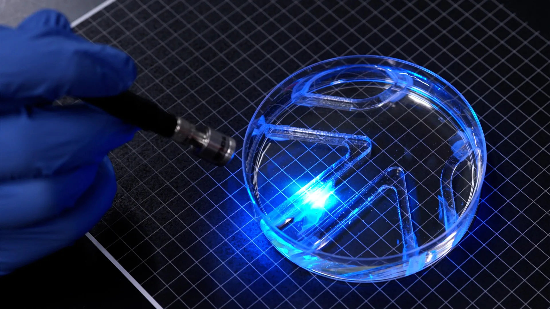
CAMBRIDGE, Mass. — In a development that bridges the gap between synthetic engineering and cellular biology, researchers at the Massachusetts Institute of Technology (MIT) have successfully designed and tested a swimming robot powered not by traditional motors, gears, or heavy mechanical parts, but by a single, meticulously engineered layer of living muscle cells.
This breakthrough, detailed in a recent study published in the journal Advanced Functional Materials, demonstrates that ultra-thin layers of biological tissue can generate the substantial mechanical force required to propel a device through fluid environments. The innovation represents a pivotal shift away from the bulky, resource-intensive designs that have historically dominated the field of biohybrid robotics, opening new doors for delicate, adaptable, and self-healing machines capable of navigating environments hostile to conventional hardware.
Main Facts
The core of the MIT innovation is an extremely thin, two-dimensional swimming robot featuring a flexible gel framework roughly the size and dimensions of a standard stick of gum. This framework bifurcates into two distinct fins, each coated with a sheet of muscle cells substantially thinner than a human hair.
To achieve remote locomotion, these living cells were genetically modified to contract reflexively when exposed to light. By selectively directing a light source onto either the left or right fin, researchers can trigger targeted muscle contractions. This causes the fins to flap rhythmically, pushing the device forward. By modulating which fin is illuminated and the frequency of the light pulses, the research team can exercise precise, real-time control over both the robot’s swimming speed and its directional heading.
In rigorous laboratory tests, the soft-bodied swimmer successfully navigated a custom-built underwater maze. At its peak operating speed, the device covered approximately four times its body length every minute. While this pace trails far behind elite human swimmers—who can achieve up to 65 body lengths per minute—it mirrors the leisurely cruising speed of a cow shark moving through the open ocean.
Funded in part by the Office of Naval Research, the project was spearheaded by a team of MIT engineers and scientists. The paper’s first author is Maheera Bawa, working alongside colleagues Arielle Berman, Laura Schwendeman, Ferdows Afghah, and Seanbiron Johnson, under the direction of Ritu Raman, associate professor of mechanical engineering at MIT.
Chronology of Development
The journey toward a muscle-powered swimming robot began years prior in Professor Raman’s laboratory, evolving through a step-by-step process of overcoming material limitations and scaling up mechanical force generation.
Phase 1: The Artificial Iris
Last year, Raman’s research group laid the groundwork for this achievement by creating an artificial muscle disk inspired by the mechanics of the human iris. For that project, the engineers patterned a soft gel disk with microscopic grooves radiating outward from the center in concentric circles. Living muscle cells were seeded onto this surface, where they naturally grew into a thin, organized layer following the pathways of the channels.
When illuminated, the muscle tissue contracted multidirectionally, causing the disk to rhythmically stretch and compress in a manner mimicking the expansion and contraction of a human pupil. While this proved that scientists could grow and control ultra-thin muscle layers in intricate geometric arrangements, the physical output was minimal. The movements were confined to a scale of roughly 100 microns—far too weak to propel an object through dense fluids like water.
Phase 2: Material Optimization
Determined to dramatically increase force production, the MIT team sought to build a more robust foundation for the living tissue. The earlier iris model had relied on fibrin, an exceptionally soft biological gel. However, the researchers discovered that fibrin degraded and shrank rapidly under the heavy mechanical loads exerted by vigorously contracting muscle cells, severely limiting effective movement.
To solve this, the engineers systematically investigated three critical "tuning knobs" of the supporting gel structure: its chemical composition, its structural stiffness, and the precise geometry of the surface grooves used to organize cellular growth. They tested various groove profiles, including narrow channels with square cross-sections and longer channels with curved bottoms. Results showed that muscle cells aligned significantly better within square-profile grooves. This alignment was vital, as cells growing in a unified direction are far more likely to fuse into coherent, synchronized muscle fibers capable of generating coordinated force.
Furthermore, the team replaced fibrin with gelatin methacrylate (GelMA), a resilient biomaterial widely utilized in tissue engineering. By fine-tuning the formulation of the GelMA, the engineers produced supporting films with varying degrees of rigidity. They discovered that stiffer gels yielded optimal results, supporting better cellular alignment and producing much stronger contractions.
Through ongoing experimentation, the team determined that a gel film measuring approximately half a millimeter thick offered the ideal equilibrium: it was sturdy enough to support a single layer of muscle cells while remaining lightweight enough to prevent the contracting tissue from shearing off the substrate.
Phase 3: Cellular "Exercise" and Assembly
Before integrating the muscle-laden gel into the final swimming robot, the researchers subjected the tissue to a regimen of biological "exercise." By administering repeated pulses of light to stimulate regular contractions, the team conditioned and strengthened the muscle fibers.
Once optimized, the gel framework was divided into two distinct fins, both patterned with square-bottomed grooves and populated with the conditioned muscle cells. The resulting device was placed in an experimental water chamber, setting the stage for successful underwater navigation trials.
Supporting Data
The development of the muscle-powered swimming robot relies on precise material metrics and biological parameters, as documented in the research team’s findings:
- Dimensions: The entire robot possesses a flexible gel framework comparable in length and width to a stick of gum.
- Thickness: The supporting gel film measures roughly 0.5 millimeters thick, providing structural integrity without excess weight. The muscle cell layer coating the fins is significantly thinner than a single human hair.
- Locomotion Speed: At maximum capacity, the robot traverses approximately four body lengths per minute.
- Comparative Kinetics: While slower than dynamic aquatic vertebrates or professional human swimmers (who achieve up to 65 body lengths per minute), the robot’s relative velocity is on par with slow-moving benthic marine life, such as the cow shark.
- Control Resolution: Independent optical targeting allows for differential fin movement, yielding precise turning radii and velocity changes via simple light-pulse frequency adjustments.
Official Responses and Perspectives
The implications of merging living biology with robotics have drawn enthusiastic responses from the academic and engineering communities, particularly regarding the philosophical and practical shift from rigid metallic machines to soft, responsive biohybrid systems.
"It takes a lot of force to move through water versus air," notes Ritu Raman, the study’s senior author and associate professor of mechanical engineering at MIT. Underscoring the efficiency of the design, she adds, "The robot’s quite strong, given its size."
Reflecting on the broader trajectory of the field, Raman contrasts their current work with prevailing methodologies in biohybrid engineering:
"Currently, biohybrid robots from our group and others’ are built from bulky, 3D chunks of lab-grown skeletal muscle that require millions of cells to fabricate. We believe that biohybrid robots powered by living muscle could one day perform delicate jobs like exploring environments too fragile or unpredictable for conventional hardware, because living tissue is soft, responsive to its surroundings, and can heal itself."
Addressing the novelty of the structural architecture, Raman highlights the leap from their previous micro-scale experiments to macroscopic aquatic locomotion:
"People hadn’t seen this muscle architecture engineered from scratch before. And the cells were moving in multiple directions. But they only moved about 100 microns. From a robotics perspective, their movements were tiny."
By successfully scaling this architecture to generate macroscopic propulsion, the team has validated a new design paradigm for soft robotics.
Implications and Future Outlook
The creation of an ultra-thin, muscle-powered swimming robot opens up expansive horizons across multiple scientific and industrial domains. By demonstrating that robust mechanical force can be harnessed from two-dimensional layers of living tissue rather than heavy motors and complex gearboxes, the MIT team has introduced a viable blueprint for a new class of biohybrid devices.
Environmental Monitoring and Exploration
Conventional underwater exploration hardware—typically constructed from rigid plastics, heavy metals, and electronic actuators—carries a high risk of damaging delicate marine ecosystems, such as coral reefs, fragile kelp forests, or sensitive deep-sea hydrothermal vents. In contrast, biohybrid robots composed largely of soft hydrogels and organic tissue present a gentle alternative. Because living tissue is inherently compliant and non-abrasive, these robots can operate in close quarters with delicate flora and fauna without causing physical trauma.
Furthermore, biological actuators are uniquely suited for aquatic environmental monitoring. Equipped with appropriate biosensors, future iterations of these robots could silently patrol vulnerable waterways, detecting chemical pollutants, monitoring water temperature fluctuations, or tracking aquatic micro-ecosystems with minimal ecological disturbance.
Medical and In Vivo Applications
Beyond open-water monitoring, the underlying technology points toward biomedical applications inside closed or sensitive environments. The ability to engineer ultra-thin, responsive muscle films that operate on light cues suggests future deployment within fluid-filled spaces inside the human body or industrial piping systems. Because biological tissue possesses self-healing properties and can interface seamlessly with organic environments, these principles could eventually guide the development of targeted drug-delivery systems or micro-surgical assistants capable of navigating complex circulatory pathways.
Next Steps for the MIT Team
Despite the success of the current maze-navigation trials, the researchers emphasize that the present device is intentionally simplistic. The primary objective of the recent study was purely foundational: to prove unequivocally that an exceptionally thin layer of engineered muscle could generate sufficient propulsive force for swimming.
With that proof-of-concept established, Raman and her colleagues are already looking toward the next iteration of their technology. "Our next goal is to optimize the body design to enable faster swimming," Raman states. By refining the hydrodynamic profile of the gel framework, improving muscle conditioning protocols, and exploring advanced optical control systems, the research team aims to dramatically increase the velocity and agility of future biohybrid swimmers.
As biohybrid engineering continues to mature, inventions like MIT’s muscle-powered swimming robot signal a blurring of boundaries between living systems and synthetic machines—paving the way for a future where technology does not just mimic nature, but actively incorporates it.